I Results Mcl
Cincinnati Bengals quarterback Joe Burrow tore his ACL and MCL in his left knee, MRI results showed on Monday.

ESPN’s Adam Schefter and Ben Baby had the news first, with NFL Network’s Ian Rapoport confirming. The former also reported there were additional “structural issues.”
Mike Garafolo of NFL Network provided some important context:
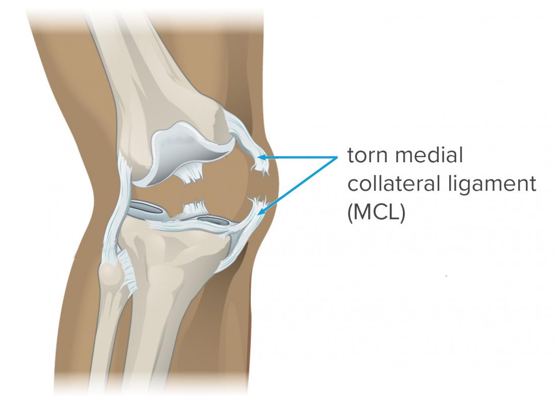
Both Stuttle and Kerlan and Glousman reported that the clinical condition referred to as MCL bursitis may result when the MCL bursa is inflamed and distended by fluid. These authors consider MCL bursitis an important cause of medial knee pain that should be differentiated from other conditions such as MCL injuries and meniscal tears. To navigate through the Ribbon, use standard browser navigation keys. To skip between groups, use Ctrl+LEFT or Ctrl+RIGHT. To jump to the first Ribbon tab use Ctrl+. Powered by JusticeTrax © 2000-2010 JusticeTrax Inc. All rights reserved. IResults by JusticeTrax Welcome! The medial collateral ligament, or MCL, of the knee can tear due to injury and cause pain. Treatment depends on the severity of the injury. Learn more about MCL tears here.
“A multi-ligament injury like Carson Wentz had a few years ago. He got hurt Dec. 10 and missed the first two games the following year. So use that as a rough guide for how long Burrow will be sidelined.”
I Results Mcl

The initial diagnosis after the injury was a torn ACL, though the report left room for possible additional damage based on MRI results.
Iresults Mcl
For his part, Burrow is playing it just like most figured he would — smooth. He offered a message to Bengals fans and those who rushed to support him, which included the likes of Russell Wilson and Patrick Mahomes.
About Ligaments and Your MCL
Mcl I Results


Ligaments are thick, strong bands of tissue that connect the ends of bones together to form a joint. Ligaments also stabilize and strengthen the joint.
Four ligaments connect the bottom of the thighbone, known as the femur, to the top of the shinbone, also known as the tibia. The ligament at the front of the knee, the anterior collateral ligament (ACL), prevents the shinbone from rotating and sliding forward off the thighbone during jumping or other activities that require quick changes in speed or direction. Immediately behind the ACL is the PCL, or posterior collateral ligament, that prevents the tibia from sliding backward of the femur.
Two ligaments, one lying on each side of the knee, provide side-to-side stability for the knee joint. The LCL, or lateral collateral ligament, is on the outer side of the knee. The MCL is on the inside of the knee, on the side of the knee that is closest to the other knee.
The ACL at the front of the knee, the PCL at the back of the knee, the LCL at the outer aspect of the knee and the MCL on the inner aspect of the knee work together to provide strength and stability to the knee. The MCL and ACL on each side of the knee work with the PCL at the back of the knee to prevent the shinbone from sliding too far backwards on the thighbone, for example.